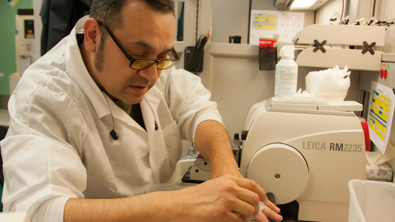
The immune-cell landscape of the human hippocampus appears to reorganize substantially beginning in midlife, according to an NIH-supported study that combined single-cell measurements across postmortem tissue from 40 adults.
The donors were neurologically healthy and ranged from age 20 to 95. Researchers focused on the hippocampus, a region central to learning and memory, and examined cells one at a time rather than averaging signals across whole tissue samples.
Resident immune cells called microglia progressively declined between roughly ages 50 and 75. At the same time, cells with stronger inflammatory signatures and features resembling immune cells derived from peripheral blood became more prominent.
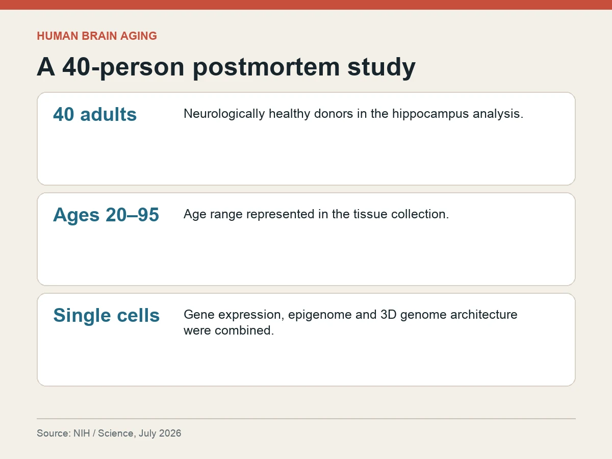
The team paired gene-expression data with the epigenome—the chemical marks that help regulate genes—and the genome’s three-dimensional architecture. That combination let researchers compare what a cell was doing with molecular clues about its identity and lineage.
Other age-linked changes appeared beyond microglia. Cells involved in maintaining the blood-brain barrier deteriorated, and multiple brain-cell types showed coordinated disruption in genome architecture and gene regulation.
The results challenge the longstanding assumption that microglia established during embryonic development simply renew themselves throughout life without a major lineage transition. They do not, however, show why the resident cells decline or where every replacement cell originates.
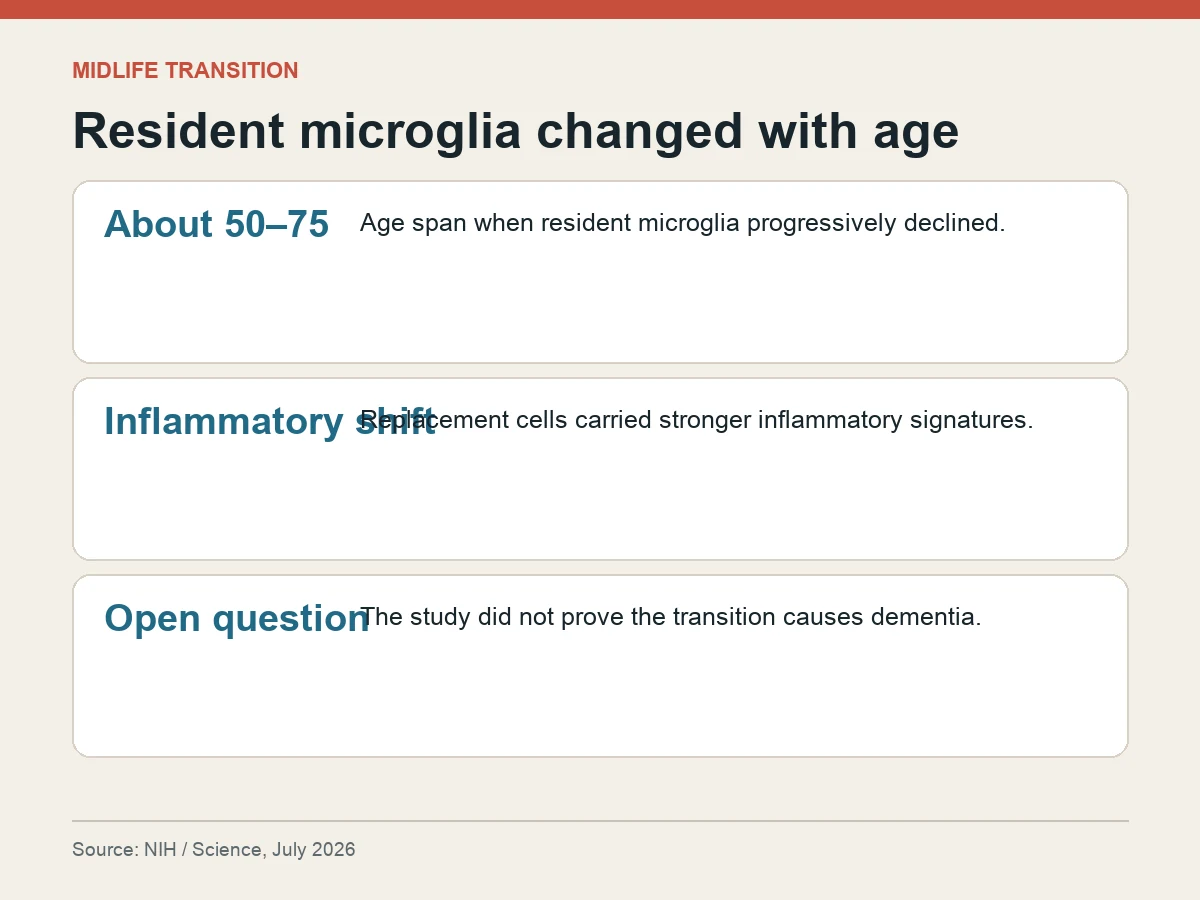
The study also does not prove a cause of dementia. Postmortem tissue provides a detailed cross-sectional comparison across ages, not a movie of cellular change within the same person, and all 40 donors were described as neurologically healthy.
Researchers plan to test whether the transition contributes directly to Alzheimer’s disease or other age-related neurological conditions. That causal step is essential before the finding can support a treatment claim or clinical recommendation.
The paper, published in Science under DOI 10.1126/science.adt8307, therefore adds a new candidate mechanism to brain aging: a midlife change in immune-cell identity accompanied by inflammatory and genomic remodeling, documented in human tissue but still awaiting disease-specific validation.